18 Differential Stains - Gram Staining
18.1 Identification of microorganisms by microscopic characterisation
The individual cells of typical bacteria are very small. To be seen clearly with a light microscope, they must be fixed to a glass slide, stained, and then examined using a microscope fitted with an oil-immersion lens (x100) which is capable of producing an overall magnification of at least x1000. (Note that the total magnification is the product of the objective and the ocular lenses – e.g., for many commonly used light microscopes, objective magnification (x100) * ocular magnification (x10) = x1000 magnification.)
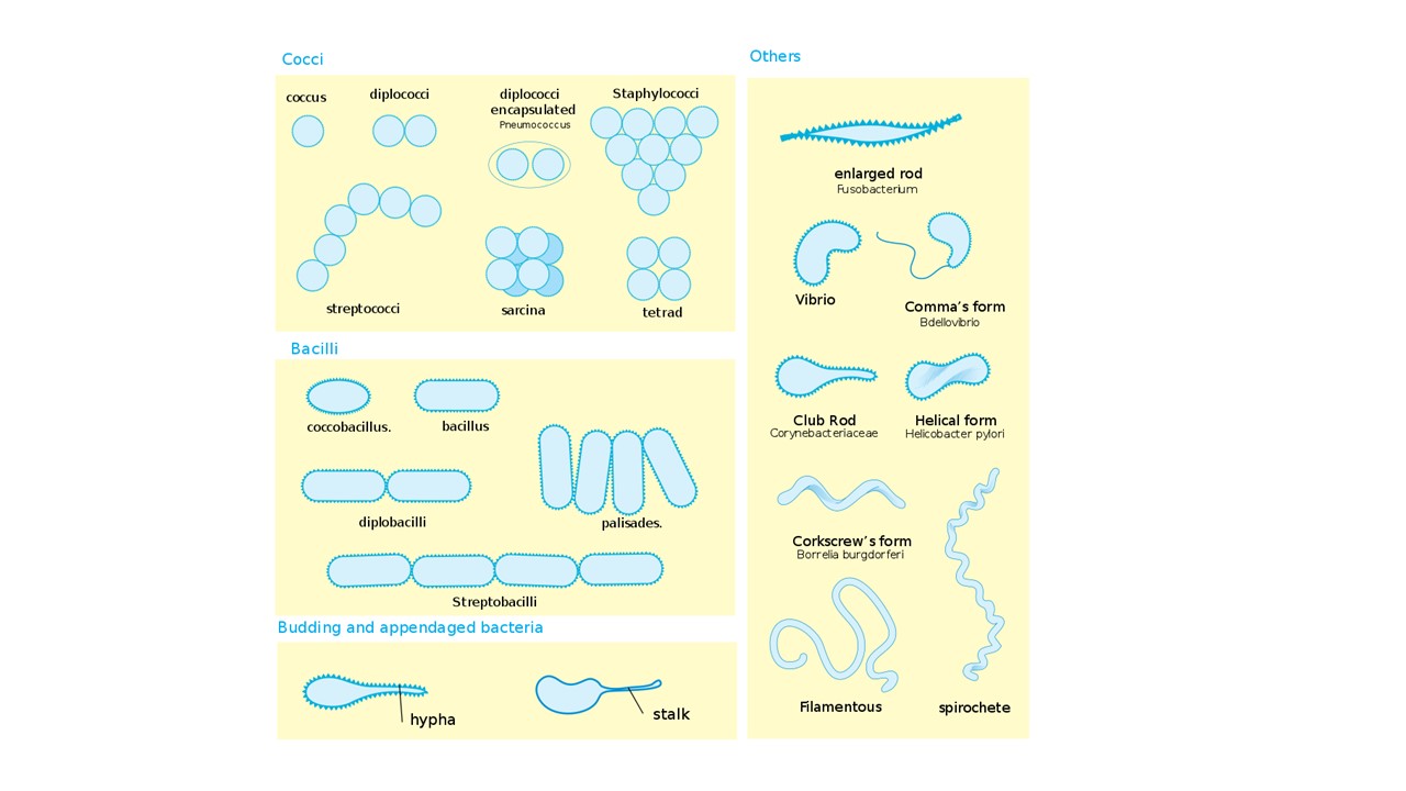
The size, shape, and arrangement of the cells (Figure 18.1) can be helpful in bacterial identification. However, this information is rarely enough to make a definitive diagnosis - there are, for example, many rod-shaped bacterial pathogens. You will need to take other information into account (often using a dichotomous key, as discussed in Section 16.2)
18.2 Differential Stains
Differential stains use multiple different chemicals which stain different cells, or cell structures, in a way which helps us to differentiate between them. Some common examples (and ones that you will see in this module) are the Gram stain and the acid-fast stain (which differentiate bacteria based on their cell wall structures).
18.3 The Gram Stain
The most common staining technique employed in microbiology is the Gram stain since it allows visualisation of the test bacterium under a light microscope and it is a discriminatory stain (i.e. it contributes to the identification of a microorganism, particularly when the information provided is used in conjunction with the macroscopic characteristics - Section 16.3).
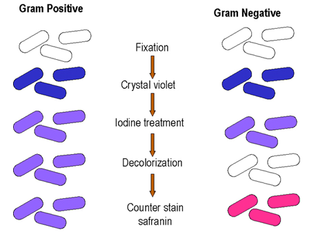
The Gram stain allows us to characterize bacteria based on the structure of their cell walls, and moreover, observing the cells under the microscope and noting their shape and arrangement (Figure 18.1) can help to narrow down a preliminary identification. In the Gram stain (Figure 18.2), test bacteria are first stained with crystal violet, which stains the cell walls of Gram positive and Gram negative bacteria. Next, iodine is added as a mordant1. The cells are then destained with alcohol and finally, counter-stained with safranin. The destaining step removes the crystal violet from Gram-negative, but not Gram-positive, cells; and therefore, only the Gram-negative cells are stained pink/red by the safranin counter-stain.
Do not put too many cells on the slide - you will need to be able to observe the cells under the microscope (which means that light must penetrate through the film of cells - if it is too thick, you won’t be able to see anything)
Follow the protocol precisely - good attention to the timing of each step is very important, you do not want to overstain or over-destain, as this can give false results.
Do not “overcook” the slide in the flame when you are fixing your bacteria - boiling the cells can change their arrangement and excessive heat can damage the cell walls
Make sure you are wearing gloves when working with the stains, and removing your gloves when working with the Bunsen burners!!!
A mordant helps the dye to bind and fixes it in place.↩︎