25 More Examples of Selective and Differential Media
25.1 Selective and Differential Media
As we have already seen, microbial growth media can be categorised as being selective or differential (Section 17.1). You have previously used nutrient agar, for growing different bacterial species, and Sabouraud/dextrose agar, for growing different fungal species - both relatively non-selective media. You have also used blood agar as a differential medium.
In this lab, we will be using two growth media that are both selective and differential: MacConkey agar, for the identification of enterobacteria, and Mannitol Salt Agar (MSA), for the identification of streptococci (to set up an experiment for our next lab).
25.1.1 MacConkey Agar
MacConkey agar (Figure 25.1) is a selective differential medium that is used for primary isolation of Enterobacteriaceae and related enteric Gram-negative bacilli and to aid in the identification of potentially pathogenic microorganisms.
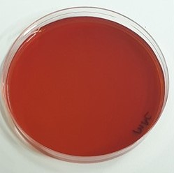
MacConkey agar is differential as it contains a sugar and the pH indicator neutral red, which can help to distinguish between bacteria which are able to metabolise the sugar, and those which cannot. It is often supplemented with lactose, but can also be supplemented with other sugars, such as sorbitol. Fermentation of the sugar in the medium produces acid, causing precipitation of the bile salts present in the medium and a change in colour due to the pH indicator. Bacteria that are not able to ferment the sugar will instead utilise the amino acids present in the medium, producing ammonia and causing the pH to increase.
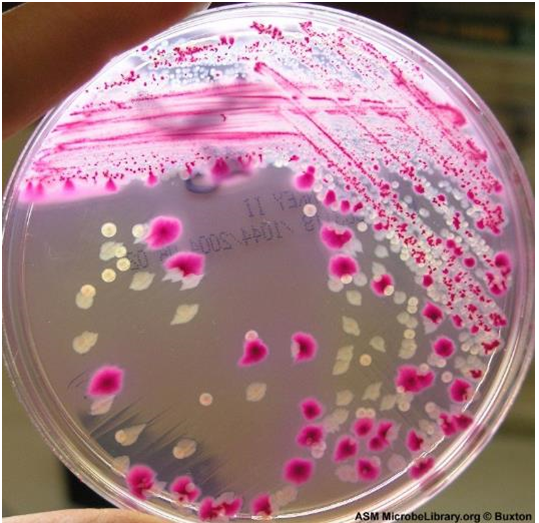
Therefore, following culture on MacConkey/lactose medium, strong lactose fermenters such as Escherichia species will produce red colonies with a surrounding zone of precipitated bile sometimes observable. The red colour is due to the indicator changing colour in response to acid production following lactose fermentation.
Slow or weak lactose fermenters such as Citrobacter and Serratia species may appear colourless after 24 hours or slightly pink after 24 - 48 hours.
Non-lactose fermenting microorganisms such as Proteus, Salmonella and Shigella species, with rare exceptions produce colourless or clear transparent colonies.
As you can see in Figure 25.2, MacConkey agar can thus be used to distinguish different bacteria based on their ability to ferment (or not) lactose.
25.1.2 Mannitol Salt Agar
Mannitol salt agar (MSA) is a selective differential medium that is used for primary isolation and identification of potentially pathogenic Staphylococcus aureus from non-pathogenic commensal microorganisms from the genus Micrococcus. MSA contains a relatively high sodium chloride content of 7.5% (w/v) which allows selection for microorganisms that can tolerate this level of sodium chloride. This makes the test selective. In addition, it contains mannitol and an indicator, phenol red. If a microorganism can ferment the mannitol, acid will be produced and the indicator will turn from red to yellow in response to the pH drop. This makes the test differential.
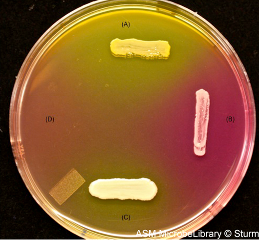
Staphylococcus aureus grows on MSA and ferments mannitol, turning the plate yellow. Staphylococcus epidermis, commonly present in the human skin flora, do not ferment mannitol, although they are capable of growth on MSA. Microbes that are not tolerant to the high concentrations of salt present in this medium (e.g., members of the genus Micrococcus such as M. luteus) cannot grow on MSA.